
Grant & I have something of an ongoing friendly competition to come up with stunning & unusual biological images. Here,via PZ (as usual!) & The Node is my latest offering:

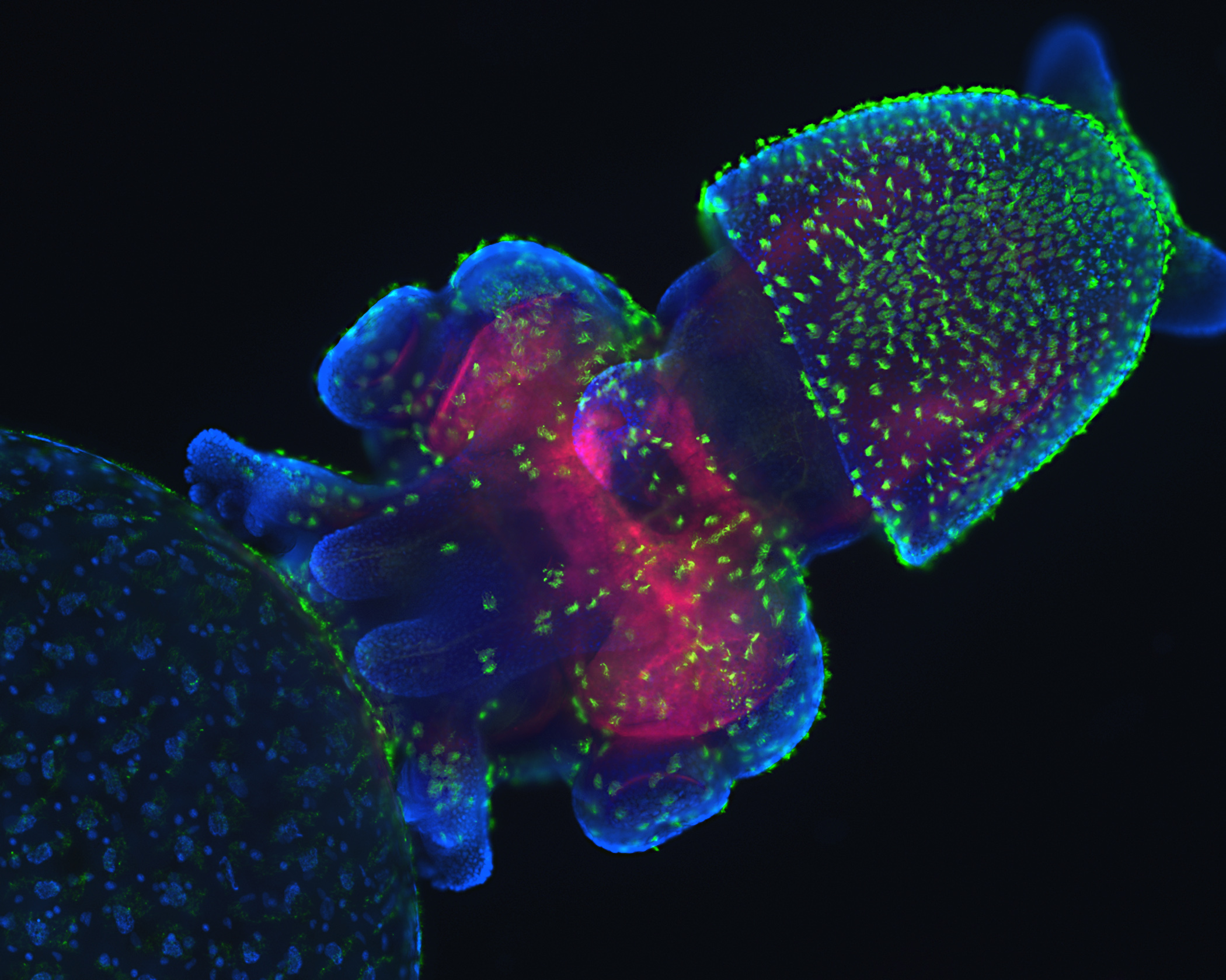
It’s a confocal microscope image of a squid embryo. The reddish areas are neural tissue (mmmm, braaaainzz) & each of those fluorescent green speckles is a tuft of cilia. (I wouldn’t have known that stuff but Sven DiMilo, one of PZ’s ‘regulars’, kindly explained it.)
Just lovely 🙂
Your turn, Grant!
Grant says:
Hey, you. You should give me a chance to find something I can beat you with, sod it 🙂
(Your pictures are way too good. Still, I’ll try…)
eviltwit says:
Wow. Makes me want to be able to breathe underwater just so I could spend weeks under the sea just enjoying colors like this.
eviltwit says:
p.s. I had to look up “confocal” and ended up on “confocal microscopy”. Glad I did. Learned something new. Cool technology!
Alison Campbell says:
A couple of years ago we had a holiday at Port Douglas (north of Cairns) & spent a day snorkelling out on the Great Barrier Reef. That was really something! Mind you, because of the way the different wavelengths of light penetrate water, once you go down more than a few metres all fish are blue 🙂
Grant says:
Eviltwit:
For whatever it’s worth, the guy who (re)invented confocal microscopy worked in the same office as me when I was a Ph.D. student.
(My reply is linked on my name for those wondering.)